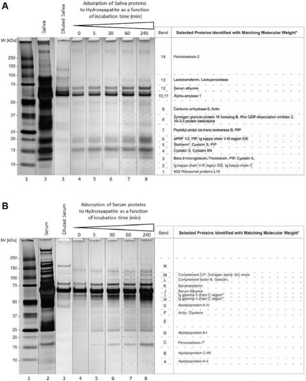
Figure 1.
Identification of proteins in saliva and serum adsorbing to hydroxyapatite. (A) Saliva pellicles and (B) serum pellicles each formed after various incubation times with hydroxyapatite. Gels were silver-stained. Lane 1, Molecular weight standard; lane 2, original saliva and serum samples (20 µL); lane 3, diluted saliva and serum samples; lanes 4 to 8, adsorbed saliva and serum fractions harvested after 0, 5, 30, 60 and 240 min incubation with hydroxyapatite (20 µL). Proteins in the excised bands were identified by mass spectrometry. Shown are proteins identified by more than 2 peptides, with theoretical molecular weights matching their apparent molecular weights in the gels (± 10 kDa; exceptions are indicated with asterisks). These proteins were among the 95% most abundant proteins identified. The proteins were excised from gels and identified by mass spectrometric analysis in 2 independent experiments.

