Figure 2.

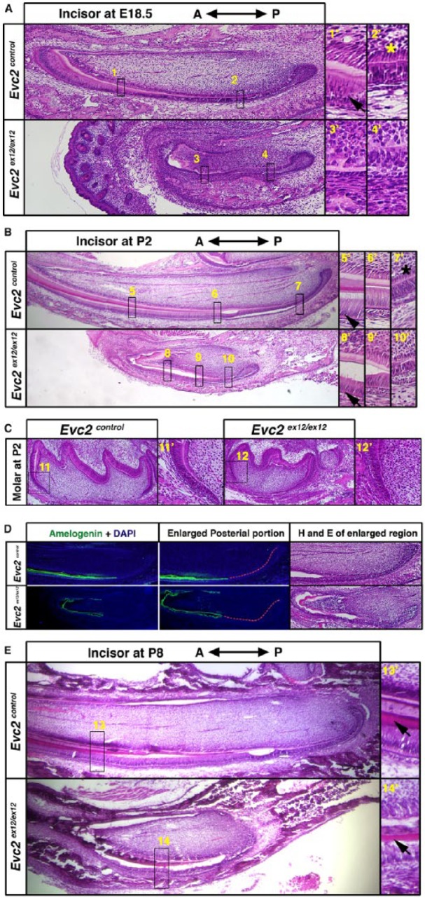
Evc2 mutant incisors showed delayed ameloblast differentiation and hypomorphic enamel formation. (A) Sagittal sections of embryonic day 18.5 (E18.5) embryonic mandible incisors. Regions in boxes in both controls and mutants were enlarged and shown on the right. Arrows indicate the ameloblasts with polarized nuclei and * indicates the odontoblasts with polarized nuclei. (B) Sagittal sections of postnatal day 2 (P2) mandible incisors. Regions in boxes in both control and mutant were enlarged and shown on the right. Arrowheads indicate ameloblasts with polarized nuclei in both controls and Evc2 mutants and * indicates the odontoblasts with polarized nuclei. (C) Sagittal sections of postnatal day 1 molar. Regions in boxes in both control and mutant were enlarged and shown. (D) Immunohistochemistry of amelogenin in mandible incisors at P2 indicates a delayed ameloblast differentiation. Posterior regions of incisors of both genotypes were enlarged and shown. Red dashed lines indicate the epithelial cells without amelogenin immunosignals. The immediate next section was processed for hematoxylin and eosin staining. (E) Sagittal sections of postnatal day 8 (P8) mandible incisors. Regions in boxes in both control and mutant were enlarged and shown on the right. Arrows indicate enamel formation in both control and Evc2 mutants.
