Figure 3.
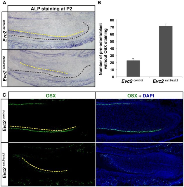
Evc2 mutant incisors showed delayed odontoblast differentiation. (A) Mandible incisors from postnatal day 2 (P2) were sectioned and processed for alkaline phosphatase (ALP) staining. Black dashed lines indicate a boundary between dental epithelium and mesenchyme. Yellow dashed lines indicate ALP-stained cells in the odontoblast layer. (B, C) Mandible incisors from P2 were sectioned and processed for immunohistochemistry for Osterix (OSX). White dashed lines separate dental epithelium and mesenchyme. Red dashed lines indicate OSX-expressing dental mesenchymal cells. Yellow dashed lines indicate OSX-stained cells in the odontoblast layer. The number of OSX-expressing dental mesenchymal cells was quantified and is shown in B (n = 4, P < 0.01).

