Figure 4.
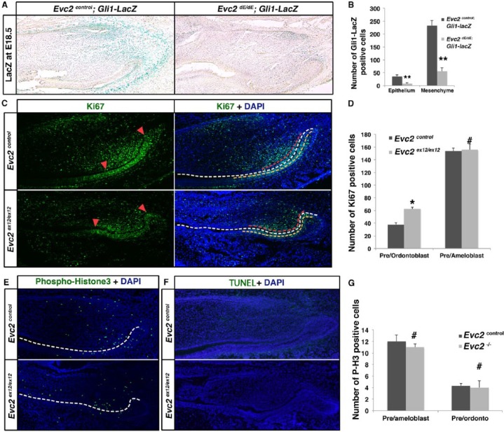
Evc2 mutant incisors showed decreased dental epithelial and mesenchymal stem cells. (A) A Gli1-LacZ reporter was introduced to label dental epithelial and mesenchymal stem cells in the incisor. E18.5 mandible incisors of indicated genotypes were processed for LacZ staining. (B) Quantification of Gli1-LacZ–positive cells in A. n = 3, **P< 0.01. (C) Immunohistochemistry of Ki-67 was applied to mark the transient amplifying (TA) cells in controls and Evc2 mutants. Red arrowheads in the left panels indicate the 2 ends of the zone of Ki-67–positive cells in the odontoblast layers. White dashed lines in the right panels indicate a boundary between dental epithelium and mesenchyme. Red dashed lines indicate areas of Ki-67–positive cells in preodontoblasts (areas between 2 red arrowheads shown in the left panels). Yellow dashed lines indicate Ki-67–positive cells in preameloblasts. (D) Quantification of Ki-67–positive cells in B. n = 4, *P < 0.05; #not significant. (E) Immunohistochemistry of phospho-histone3 (P-H3) depicts proliferating cells in control and Evc2 mutant incisors at postnatal day 2 (P2). White dashed line indicates a boundary between dental epithelium and dental mesenchyme. (F) Terminal deoxynucleotidyl transferase dUTP nick end labeling (TUNEL) staining indicates apoptotic cells in control and Evc2 mutant incisor at P2. (G) Quantification of P-H3–positive cells in C. n = 4, #not significant.

