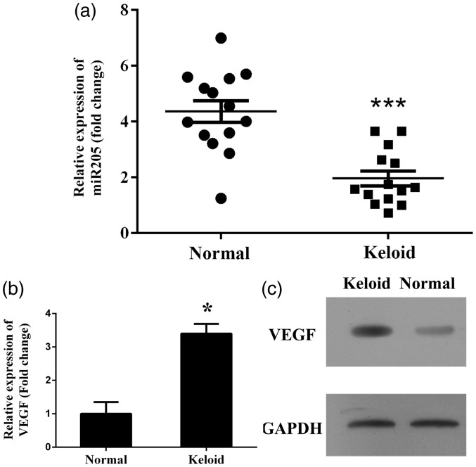
Figure 1.
Investigation of expression status of miR-205-5p and VEGF in clinical keloids and HKF. (a) Quantitative analysis results of miR-205-5p levels as detected by RT-qPCR, levels of miR-205-5p were reduced in keloid tissues compared to normal tissues. (b) Representative analysis results of VEGF levels as detected by RT-qPCR, mRNA level of VEGF was increased in keloid tissues compared to normal tissues. “*”, P < 0.05 compared with normal tissues or cells. “***”, P < 0.001 compared with normal tissues or cells. (c) Representative image of protein level of VEGF as detected by western blotting assay, protein level of VEGF was increased in keloid tissues compared to normal tissues

