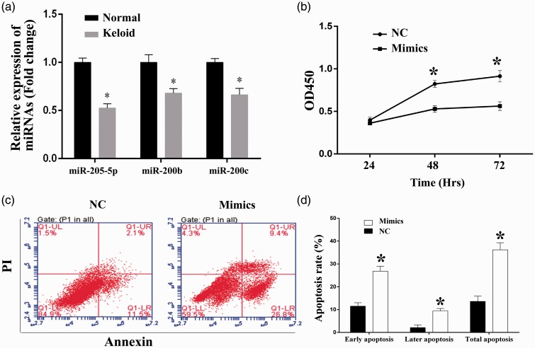
Figure 2.
Overexpression of miR-205-5p impaired the cell viability, induced cell apoptosis, and reduced the cell invasion and migration ability in HKF. (a) Quantitative analysis results of levels of miR-205-5p, miR200b, and miR200c as detected by RT-qPCR, mRNA levels were reduced in HKF compared to normal skin cells. (b) Quantitative analysis results of CCK-8 assay, transfection of miR-205-5p mimics inhibited the cell proliferation in HKF. (c) Representative images of cell apoptosis as detected by flow cytometry, transfection of miR-205-5p mimics induced cell apoptosis in HKF. (d) The percentage of early, late phase, and total apoptosis cells of each group. “*”, P < 0.05 compared with NC group. LR (lower right): Early apoptosis cells, UR (upper right): Later phase apoptosis cells, LL (lower left): Viable cells, UL (upper left): Necrosis cells. (A color version of this figure is available in the online journal.)

