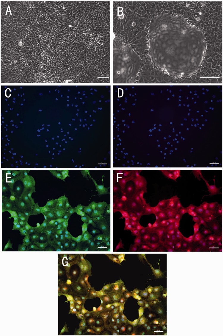
Figure 2.
Representative images of light microscopic observation and immuno-fluorescence staining of primary cultured porcine thyroid cells. (a) The monolayer porcine thyroid cells in culture after two days of incubation. (b) The thyroid cells with the hemisphere-like domes formed in culture after four days of incubation. (a and b) Scale bar = 100 µm. (c) Negative control for antirabbit IgG. (d) Negative control for antigoat IgG. (e) Positive staining of sodium-iodide symporter (NIS). (f) Positive staining of thyroglobulin (Tg). (g) Composite picture of NIS, Tg, and DAPI. (c to g) Scale bar = 50 µm. (A color version of this figure is available in the online journal.)

