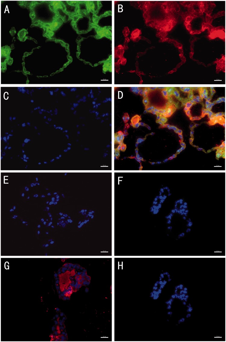
Figure 8.
Representative images of immuno-fluorescence staining of the microbeads at day 20. The images show that there were cell follicular structures inside the microbeads, which stained positive for both sodium-iodide symporter (NIS) (a) and thyroglobulin (Tg) (b). Cells were stained with DAPI only (c). Composite image including NIS, Tg and DAPI (d). Negative control of antigoat IgG (e). Negative control of antirabbit IgG (f). Thyroxine was detected in the lumen of follicular spheres (g). Negative controls of antimouse IgG (h). Scale bars on the images depict 20 µm. (A color version of this figure is available in the online journal.)

