
Figure 1.
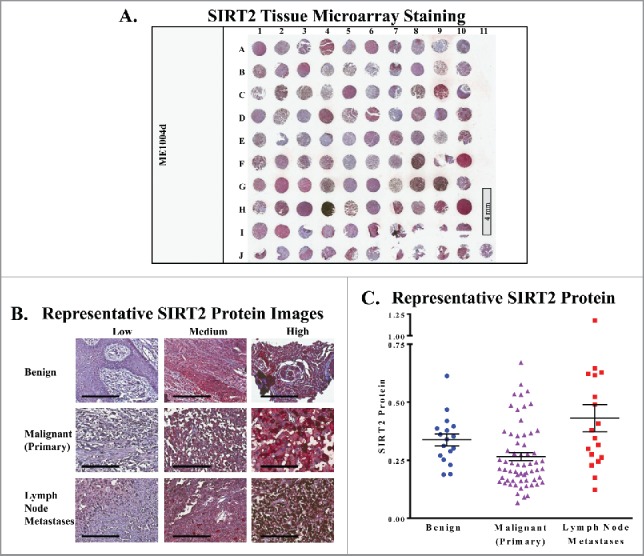
SIRT2 staining is increased in human metastatic melanoma samples. (A) Whole image of the SIRT2/AEC stained slide with hematoxylin counterstaining. ME1004d indicates the catalog number from US Biomax, Inc. that was used for the study. (B) Representative 20x images of individual cores with low, medium, and high SIRT2 expression are shown. Scale bar = 200 μm. (C) The mean whole cell SIRT2 protein levels of each core as determined by Vectra analysis are plotted. Error bars represent mean ± SEM.