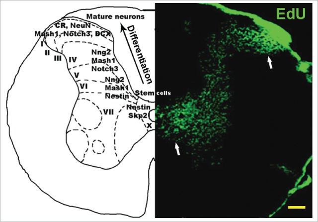
Figure 1.

Neural stem cells are continuously generated in normal mouse spinal cord. Right panel: 3-month old mouse spinal cord stained with EdU, which labels proliferating cells. Higher density EdU staining is present around the central canal (lamina X), where neural stem cells are generated from the ependymal layer, and in laminae I/II, where these cells accumulate after migrating along the dorsal horn. Left panel: Distribution of neural progenitor markers across spinal cord layers. The ependymal cell layer stains for stem cell markers Skp2 and Notch2. As neural stem cells migrate from lamina X toward lamina I, neural markers are expressed sequentially, including nestin, Mash1, Ngn2, Notch3, DCX, CR and NeuN, showing increasing levels of neuronal differentiation toward the peripheral dorsal horn. CR = calretinin, DCX = doublecortin. Scale bar: 1 mm.
