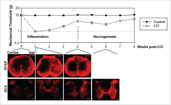
Figure 2.

Allodynia has 2 stages, corresponding to inflammation and neurogenesis. Upper panel: Nociceptive sensitivity measured after sciatic nerve ligation in rats (the CCI model of chronic pain), shows 2 distinct stages when represented on a semi-log scale. First stage, lasting approximately 4 weeks after nerve injury, corresponds to inflammatory pain, reflected in the activation of microglia (not shown) and reactive astrocytes, identified by GFAP staining. GFAP staining ipsilateral to nerve injury (Ipsi) peaks at 1 week after nerve injury, then decays back to normal by 4 weeks, mirroring the first stage of allodynia. The second stage of allodynia, lasting from week 4 to weeks 10-12 post-CCI, corresponds to an increase in immature neuron markers such as doublecortin (DCX) and Mash1 (not shown), predominantly on the ipsilateral side. The 4-week delay is explained by the time necessary for neural progenitor cells' proliferation in lamina X, migration to laminae I/II, and partial differentiation into immature neurons. The buildup of highly excitable immature neurons in nociceptive laminae I/II generates a second peak of nociceptive sensitivity that correlates with the peak in the expression of immature neuron markers. Scale bars: 1 mm.
