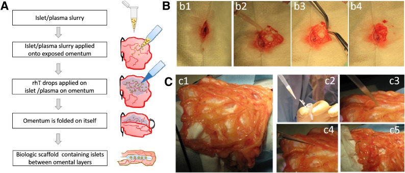
Figure 1.
Intraomental islet implantation within a biologic scaffold. A: Schematic diagram of the transplant procedure. B: Procedure in rat. C: Procedure in NHP. After midline laparotomy (b1), the omentum is gently exteriorized and opened (b2 and c1). The islet graft, resuspended in autologous plasma (c2), is gently distributed onto the omentum (b3 and c3). Recombinant human thrombin is added onto the islets on the omental surface to induce gel formation (c4), and then the omentum is folded to increase the contact of the graft to the vascularized omentum (b4 and c5). Nonresorbable stitches were placed on the far outer margins of the graft in the NHP (c5) for easier identification of the graft area at the time of graft removal.

