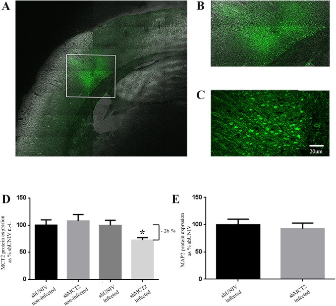
Fig 1. Characterization of the neuronal MCT2 knockdown in the rat somatosensory cortex following the injection of a lentiviral vector to selectively express a shRNA against MCT2.
(A, B and C) Fluorescence signal from the expressed Green Fluorescent Protein (GFP) on a coronal section of a rat injected with a lentiviral vector expressing GFP as a marker of infected cells. (A) represents a mosaic of pictures taken at a magnification of 20X with the tilescan function of the confocal microscope. (B) represents a zoom of the area on the mosaic delineated with a white frame (C) represents a picture taken at a magnification of 40X with the confocal microscope. (D) Quantification of the MCT2 immunofluorescence in neurons of the rat somatosensory cortex infected with either a shRNA against MCT2 (shMCT2) or a control shRNA (shUNIV), or non-infected from the same respective sections. The MCT2 immunofluorescence average value for non-infected neurons in sections injected with the shUNIV lentiviral vector was set at 100%. (E) Quantification of the MAP2 immunofluorescence in neurons of the rat somatosensory cortex infected with either a shRNA against MCT2 (shMCT2) or a control shRNA (shUNIV). Data represent mean ± SEM of a total of 27 neurons from three sections for each condition and were statistically analyzed with a Student t-test with Welch’s correction. *p < 0.05 vs. shUNIV infected.

