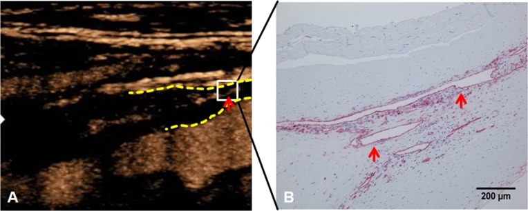
Fig 4. Neovascularization on contrast-enhanced ultrasound (CEUS) and immunohistochemistry.
(A) Contrast-enhanced ultrasound (CEUS) of carotid stenosis. The dotted yellow lines mark the proximal beginning of the carotid plaque. The red arrow marks an area of focal neovascularization. (B) The area corresponding to the white square in A was further analyzed with anti-cd31 immunohistochemistry. Note pronounced neovascularization (red arrows) in the surgical specimen.

