Figure 3.
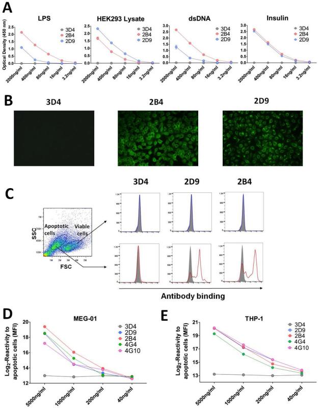
(A) Reactivity of cell culture supernatants to multiple antigens. The reactivity of polyreactive antibodies 2B4 and 2D9 and non-polyreactive antibody 3D4 to LPS, dsDNA, insulin and HEK293 lysate was assessed by ELISA using an anti-IgM secondary antibody. (B) Reactivity of cell culture supernatants to Hep-2 cells. Staining of Hep-2 cells was carried out with polyreactive clones 2B4 and 2D9 and control non-polyreactive clone 3D4. Staining was revealed using an anti-IgM secondary antibody conjugated to fluorescein isothiocyanate. All pictures were acquired using the same settings for consistency. (C) Reactivity of cell culture supernatants to Jurkat cells. Cell culture supernatants from polyreactive clones 2B4 and 2D9 as well as control non-polyreactive clone 3D4, were used to stain a mixture of viable and apoptotic Jurkat cells. Staining was revealed using an anti-IgM secondary antibody. Results are reported after gating on viable cells (upper panels) or apoptotic cells (lower panels). Filled gray histograms correspond to the signal generated by staining with the secondary antibody alone. (D) Reactivity of cell culture supernatants to apoptotic MEG-01 cell line. Cell culture supernatants at different concentration from polyreactive clones 2B4, 2D9, 4G4 and 4G10 as well as control non-polyreactive clone 3D4, were used to stain apoptotic MEG-01. Staining was revealed using an anti-IgM secondary antibody. Reactivity is reported as Log2 values of MFI. (E) Reactivity of cell culture supernatants to apoptotic THP-1 cell line. Cell culture supernatants at different concentration from polyreactive clones 2B4, 2D9, 4G4 and 4G10 as well as control non-polyreactive clone 3D4, were used to stain apoptotic THP-1 cell line. Staining was revealed using an anti-IgM secondary antibody. Reactivity is reported as Log2 values of MFI.

