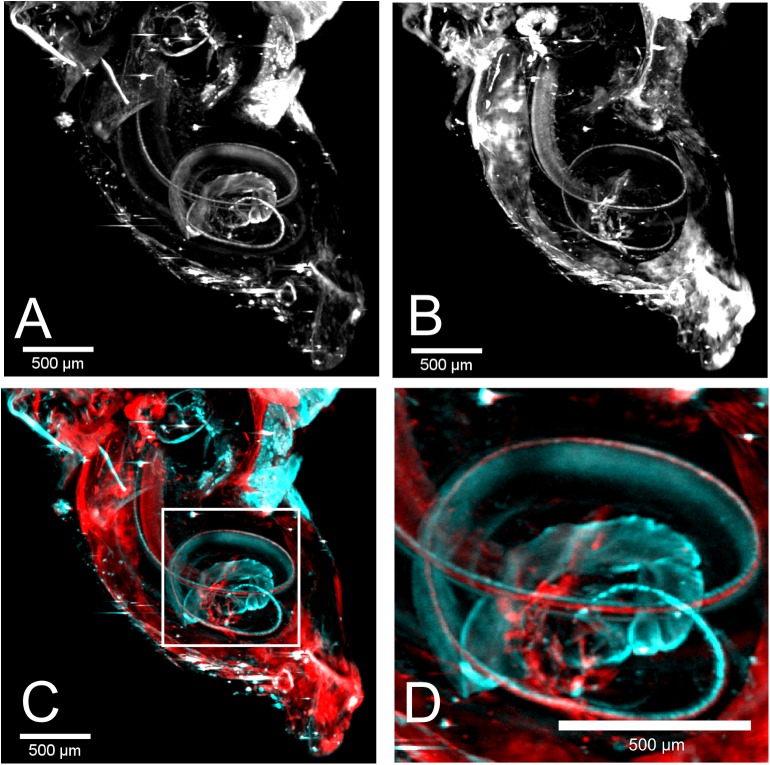
Fig 4. Double labeling of cochlea 3 with a combination of two antibodies.
(A) The MIP of the neurofilament excited at 520 nm is shown. The helical shape is visible as for sample 1. (B) The labeled inner hair cells also appear in a helical shape in the MIP excited at 635 nm. (C) An overlay of neurofilament (cyan) and hair cells (red) is shown. (D) Higher magnification of the highlighted region in C. Due to the overlay the arrangement of neurofilament and hair cells becomes visible.

