
Fig. 5. β structure of the kinase core is anchored to the hydrophobic core and recruited for catalysis by ATP binding.
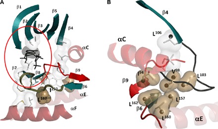
(A) The β sheet of the N-lobe (β1 to β5) is anchored to the adenine ring of ATP through V57 (β2) and A70 (β3). In the C-lobe, β7 and β8 are anchored to the adenine ring through L173 (β7). The catalytic loop that spans β6 and β7 is anchored to the F helix by L167; the Mg loop (red) bridges β8 and β9. (B) Another conserved structural element in the N-lobe is the αC-β4 loop that is characterized by a stable β turn. This loop also contains many key residues with highly correlated chemical shift changes (black spheres).