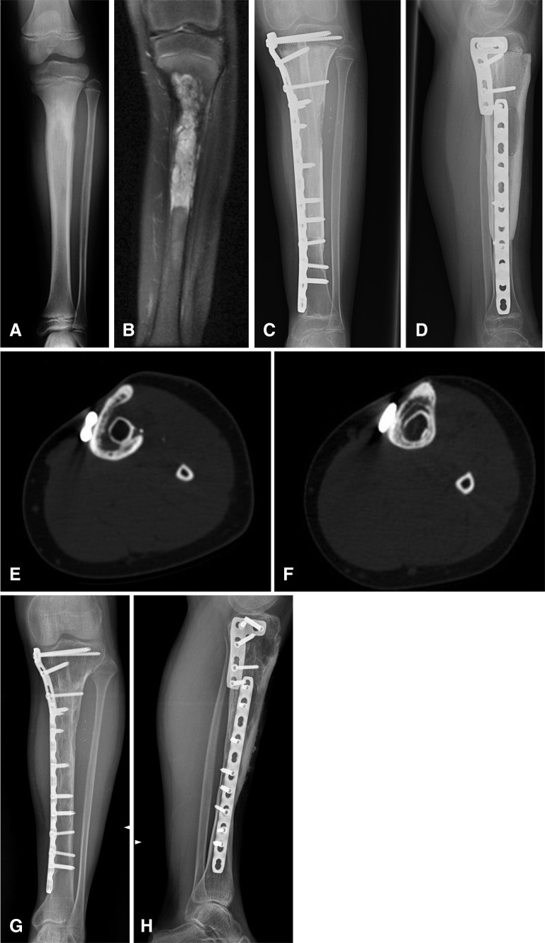
Fig. 1A–H.
(A) A plain radiograph shows a Ewing’s sarcoma of the proximal tibia diaphysis in an 11-year-old girl. (B) A representative coronal preoperative MR image of the tibia is shown. (C) AP and (D) lateral postoperative radiographs show the appearance 1 year after free vascularized fibula and massive bone allograft reconstruction. The oval slot for the fibula pedicle is evident on the lateral view radiograph. (E) An axial CT section shows the bony bridge between the inlaid fibula and allograft indicating progressive fusion at 4 years followup. (F) This axial CT section shows gradual incorporation of the free vascularized fibula with massive bone allograft at 9 years followup. (G) AP and (H) lateral radiographs obtained at the 10-year followup show the appearance of the free vascularized fibula with massive bone allograft. Incorporation of the vascularized fibula graft with allograft is observed by an indistinct appearance of the fibula graft.

