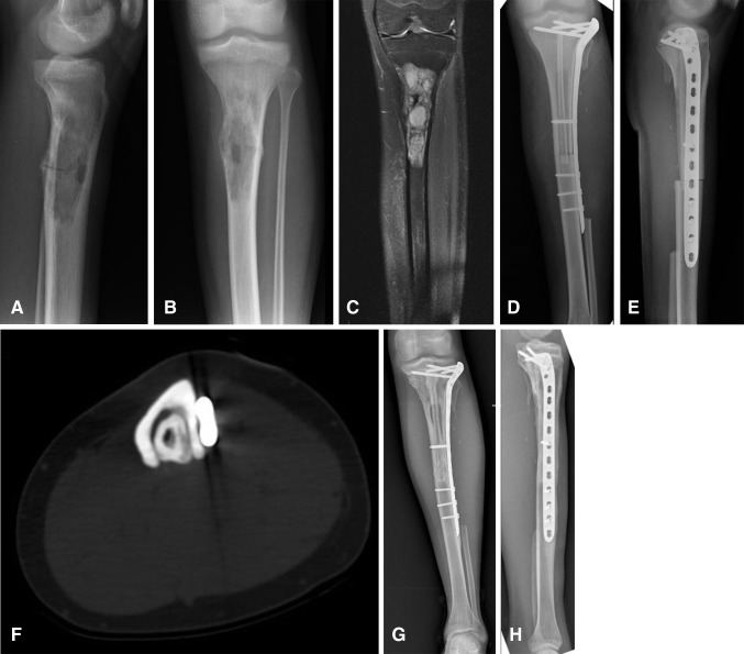
Fig. 2A–H.
(A) Lateral and (B) AP plain radiographs show the proximal tibia diaphysis involvement in a 17-year-old girl with osteosarcoma. (C) A coronal preoperative MR image of the tibia shows the extent of the tumor in the medullary cavity. Immediate postoperative (D) AP and (E) lateral view radiographs show the pedicled vascularized fibula and massive bone allograft construct. A long cortical window (lucent area in diaphysis) in the allograft to accommodate the pedicled fibula graft is seen on the AP view. (F) An axial CT section shows gradual hypertrophy of the pedicled vascularized fibula inside the massive bone allograft at 3 years followup. (G) AP and (H) lateral view radiographs show the progressive incorporation of the pedicled fibula graft with allograft at 8 years postoperatively.

