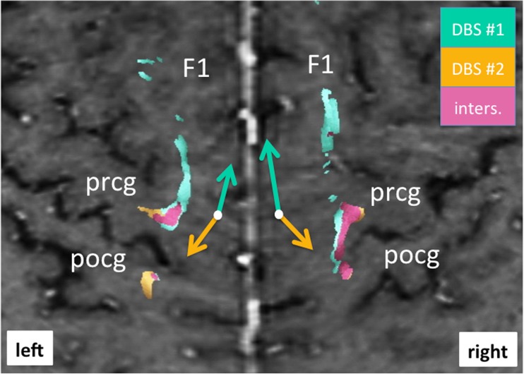
Fig. 4.
Bilateral curved surface or Mercator projection showing the cortical connections of fiber tracts involved with DBS. Note how revision surgery (DBS#2, yellow) shows a more posterior and lateral cortical connection (yellow arrow) focusing more on the precentral gyrus (prcg) and (to a lesser extent) postcentral gyrus (pcog). Projections form the initial DBS surgery (DBS#1, green) project more anteriorly and medially (as indicated by green arrows) (for legend cf. Fig. 3)

