Figure 3.
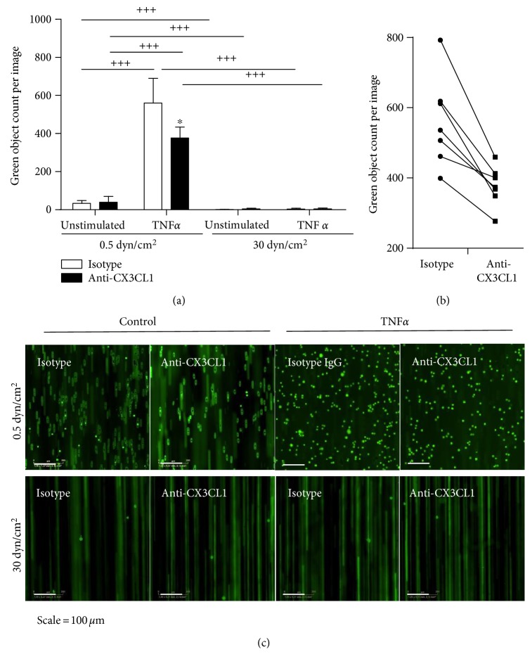
Regulation of CX3CL1 dependent THP-1 cell adhesion by shear stress. (a–c) HUVECs were cultured for 24 h and subsequently stimulated with or without TNFα for 24 h at the indicated levels of shear stress. Cells were then treated with a neutralizing antibody against CX3CL1 or an IgG isotype control for 0.5 h. Subsequently, fluorescently labelled THP-1 cells were perfused over the endothelial cell layer. THP-1 cells were visualized by fluorescence microscopy, and for each experiment adhered, THP-1 cells were counted in at least 8 images (a). Data are shown as mean + SD of 3–8 experiments as indicated. Statistical differences in comparison to the IgG isotype control or the specified flow conditions are indicated by asterisks (∗p ≤ 0.05) or crosses (+++p ≤ 0.001), respectively. (b) The effect of the inhibitory antibody against CX3CL1 is shown for each experiment performed with a different endothelial isolate. (c) Representative images showing adherent THP-1 cells (green bright circular objects) and flowing THP-1 cells (green lines) for all conditions.

