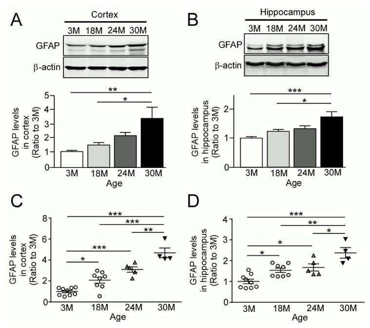
Fig. 1.

Levels of the astrocyte marker GFAP are increased in an age-dependent manner. (A and B) GFAP levels were analyzed in the cortex (A) and hippocampus (B) from wild-type C57BL/6 mice by Western blotting at the ages of 3, 18, 24 and 30 months (n=4/group). (C and D) GFAP levels were analyzed in the cortex (C) and hippocampus (D) from the above described mice by ELISA (n=4–9/group). Data are plotted as mean ± SEM. *p<0.05, **p<0.01, ***p<0.001.
