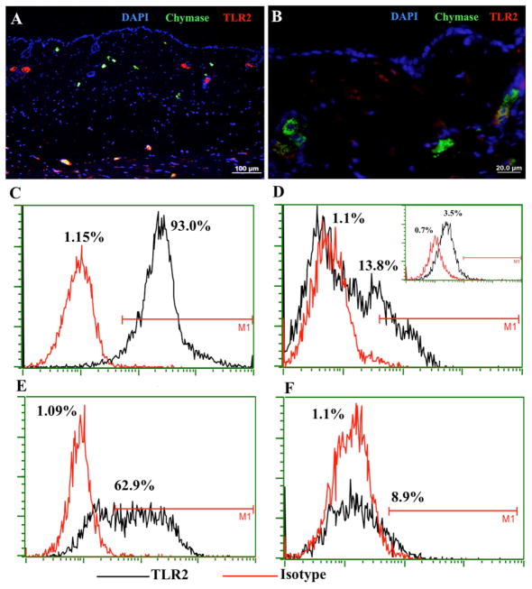
Figure 3. TLR2 MC expression in the skin.
(A) SPF mouse skin was stained with anti-chymase (green), anti-TLR2 (red), and DAPI (blue) to define TLR2 presence on dermal MCs. TLR2 is visible in the deeper layer of dermis (yellow cells at the bottom) and green cells indicate MCs that do not express TLR2; (B) Magnification of chymase positive MCs that do not express TLR2. (C–D) FACS plots and enumeration of murine bone marrow-derived MCs that were differentiated in vitro without (C) or with (D) dermal fibroblasts for 7 days, stained with anti-TLR2 antibody, and then analyzed by FACS. The inset in (D) shows the expression of TLR2 in MCs derived from the bone marrow of Tlr2−/− mice; (E–F) Human CD34+ cord blood cell-derived MCs were differentiated in vitro without (E) or with (F) fibroblasts for 7 days, stained with anti-TLR2 antibody, and then analyzed by FACS.

