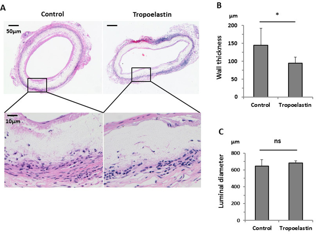
Fig. 2.
Histology and morphometric data of control and tropoelastin-seeded bioresorbable arterial vascular grafts. (A) H&E staining showed cell infiltration within the scaffolds, in both groups, 8 weeks after implantation. (B) Tropoelastin significantly decreased the wall thickness of the graft. (C) There was no significant difference between groups in the luminal diameter. *: P < 0.05, ns: no significant difference.

