Fig. 5.
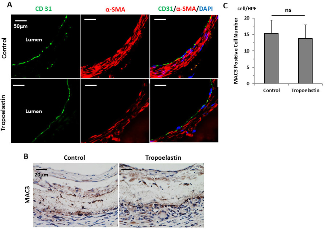
Endothelialization and macrophage infiltration of control and tropoelastin seeded bioresorbable arterial vascular grafts. (A) Representative immunofluorescent images of endothelial cells (CD31) and smooth muscle cells (α-SMA). A layer of endothelial cell coverage on the luminal side of the scaffolds is surrounded by a SMC layer in both groups equally. (B) Representative immunohistochemical images for MAC3. (C) MAC3 levels for the two groups were indistinguishable. HPF: high power field. ns: no significant difference.

