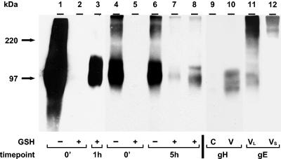
FIG. 4.
Incorporation of endocytosed gH into VZV virions. Virions were isolated as described in the legend to Fig. 3 (lanes 8 and 10). Cell lysates were collected before the isolation of virions (lanes 7 and 9). Immunoprecipitated gH samples (lanes 1 to 8) or samples without immunoprecipitation (lanes 9 and 10) were separated by SDS-8% PAGE under nonreducing conditions. Biotinylated glycoproteins were detected with Streptavidin-HRP (lanes 1 to 8). In lanes 9 and 10, Western blotting was performed with an anti-gH MAb. To detect gH-gE complex formation, the nitrocellulose membrane from lane 8 was stripped and probed for a second time with a mouse anti-gE MAb (lane 11). Lane 12 represents a shorter exposure of lane 11 to confirm the migration of the higher-molecular-mass form of gE. VL and VS indicate longer and shorter exposures of the virion sample, respectively. Lanes 1 to 3, lanes 4 to 8, and lanes 9 and 10 represent three separate gels. The corresponding molecular mass markers are indicated on the left.

