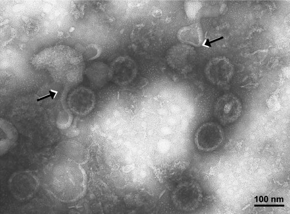
FIG. 6.
Virion fraction from the second sedimentation gradient. The virion fraction was processed for TEM as described in the text. Enveloped and nonenveloped particles were easily distinguished. Capsid morphologies were previously described (26). Partially unenveloped virions were seen; in these virions, the envelope had detached from its nucleocapsid (arrows), most likely during centrifugation in two KT-glycerol gradients. The sample was viewed at an accelerating voltage of 75,000 V and at a magnification of ×80,000.

