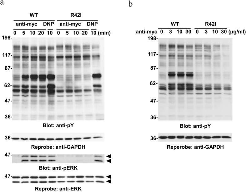
Figure 2. Engagement of Mincle induces protein tyrosine phosphorylation and ERK phosphorylation in RBL-2H3 cells.
(a) Time course. Cell lines expressing WT Mincle or the R42I mutant were stimulated with or without 10 μg/ml anti-myc mAb (anti-myc) for the indicated periods of time or preincubated overnight with anti-DNP IgE mAb and then stimulated with 300 ng/ml DNP-BSA for 10 min (DNP). (b) Dose dependency. Cell lines expressing WT Mincle or the R42I mutant were stimulated with the indicated concentrations of the anti-myc mAb for 30 min. (a and b) Detergent-soluble lysates were analyzed by immunoblotting with the indicated antibodies. Molecular size markers are indicated at the left in kDa. Data are representative of three independent experiments using PA-11 (WT) and R42I-3 (R42I) cell lines. Similar results were obtained from the other cloned cell lines.

