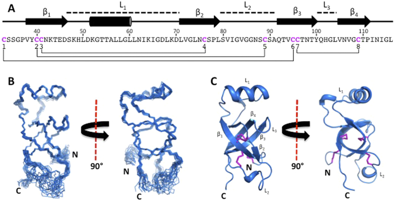
Figure 2. Solution structure of SC16.
(A) The sequence of SC16 from the first cysteine residue to the C-terminus. The secondary structure is indicated for each residue and the locations of the loops, β-sheets and helix of SC16 are indicated. Cysteine residues are coloured fuchsia and disulphide linkages are indicated by a line. (B) Superposition of the 20 lowest-energy structures of SC16, with only the N, Cα, and C’ atoms shown. (C) A ribbon diagram of the lowest-energy SC16 structure. Disulphide bonds are shown in fuchsia and the β-sheets and loops are indicated.

