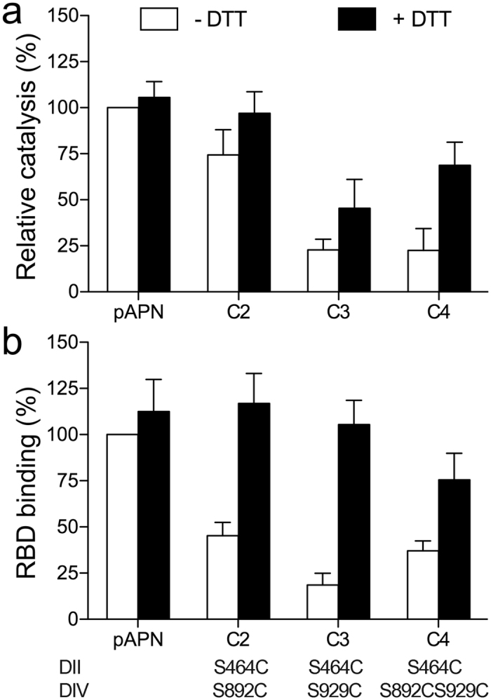
Figure 6. Locking the pAPN closed form with disulfide bonds inhibits its functions.

Relative pAPN catalysis (a) and TGEV RBD binding (b) activity of native or pAPN-cysteine mutants (C2-C4) expressed on the 293T cell surface. Cells were incubated alone or with DTT (5 mM; 30 min, 37 °C) before assays. Values expressed as a percentage of wild type pAPN values in the absence of DTT. Activities were normalized to the amount of cell surface protein expressed as monitored by flow cytometry (see Methods). Catalysis was determined at 60 min as absorbance at OD405 nm (see Fig. 3 and Methods). Relative RBD-Fc binding to transfected cells determined from mean fluorescence intensity computed by flow cytometry as in Fig. 4c. Domain II and IV residues replaced by cysteine are indicated at bottom (see Supplementary Fig. S4). Mean ± SD (n ≥ 5).
