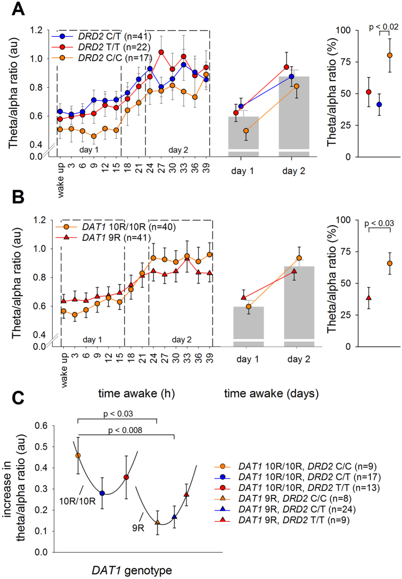
Figure 3. The neurophysiological consequences of sleep deprivation split by the DAT1-DRD2 combined genotypes.
Effect of 40 hours prolonged wakefulness on the increase in EEG theta-to-alpha ratio (TAR), split by DAT1, DRD2, and combined DAT1-DRD2 genotypes, all showing significant genotype x sleep deprivation interactions (qFDR < 0.02). The portion of the waking EEG recordings with eyes open was analyzed. (A,B) Left: TAR quantified at 3-hour intervals for DAT1 (panel A; 10R/10R: orange, 9R: red) and DRD2 (panel B; C/C: orange, C/T: blue, T/T: red) genotypes. Right: Change in TAR from day 1 to day 2 in DAT1 and DRD2 genotypes. Statistics revealed a significant effect of sleep deprivation on TAR, which was stronger in 10R/10R homozygotes than 9R-allele carriers of DAT1 and in DRD2 C/C homozygotes compared to C/T heterozygotes. (C) Increase in TAR from day 1 to day 2 in the combined DAT1-DRD2 genotype groups. The sleep deprivation-induced increase in TAR is described by a U-shaped curve with an arbitrary horizontal axis, split by DAT1 genotype. P-values represent the least significant difference (LSD) following the corresponding ANOVA.

