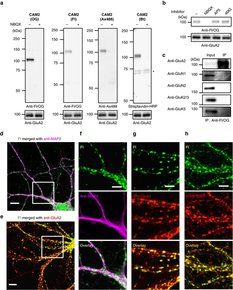
Figure 3. Chemical labelling of native AMPARs in cultured neurons.
(a) Western blot analyses of cultured neurons after labelling using CAM reagents. Cultured cortical neurons were treated with 1 μM of CAM2(OG), CAM2(Fl), CAM2(Ax488), or CAM2(Bt) in the absence or presence of 10 μM NBQX in serum free Neurobasal medium. The cell lysates were analyzed by western blot using anti-Fl/OG, anti-Ax488, or anti-GluA2 antibody, or by biotin blotting using streptavidin-HRP. * indicates biotinylated proteins endogenously expressed in the neurons. (b) Effect of competitive antagonists for glutamate receptors on chemical labelling of native AMPARs in cultured neurons. Western blot analyses of cultured neurons after labelling using CAM reagents are shown. Cultured cortical neurons were treated with 1 μM of CAM2(OG) in the absence or presence of 10 μM NBQX, 10 μM AP5, or 10 μM (2S,4R)-4-methyl glutamate (4MG) to examine selective labelling of AMPARs among the ionotropic glutamate receptor family. (c) Analyses of labelled proteins in cultured neurons by immunoprecipitation using anti-Fl/OG antibody. Chemical labelling was conducted with the same procedure described in a. After lysis of labelled cultured neurons by CAM2(Fl), the cell lysate was immunoprecipitated with anti-Fl/OG antibodies. The immunoprecipitates were analyzed by western blot using glutamate receptor-specific antibodies. (d–h) Confocal imaging of cultured neurons after labelling using CAM reagents. Cultured hippocampal neurons labelled with 1 μM CAM2(Fl) were fixed, permeabilized and immunostained using anti-MAP2 (in d,f), anti-GluA2 (in e,g) or anti-PSD95 antibody (in h). White square ROIs indicated in d,e are expanded in f,g, respectively. Scale bars, 10 μm (d,e) and 5 μm (f–h). Full blots for b and c are shown in Supplementary Fig. 22.

