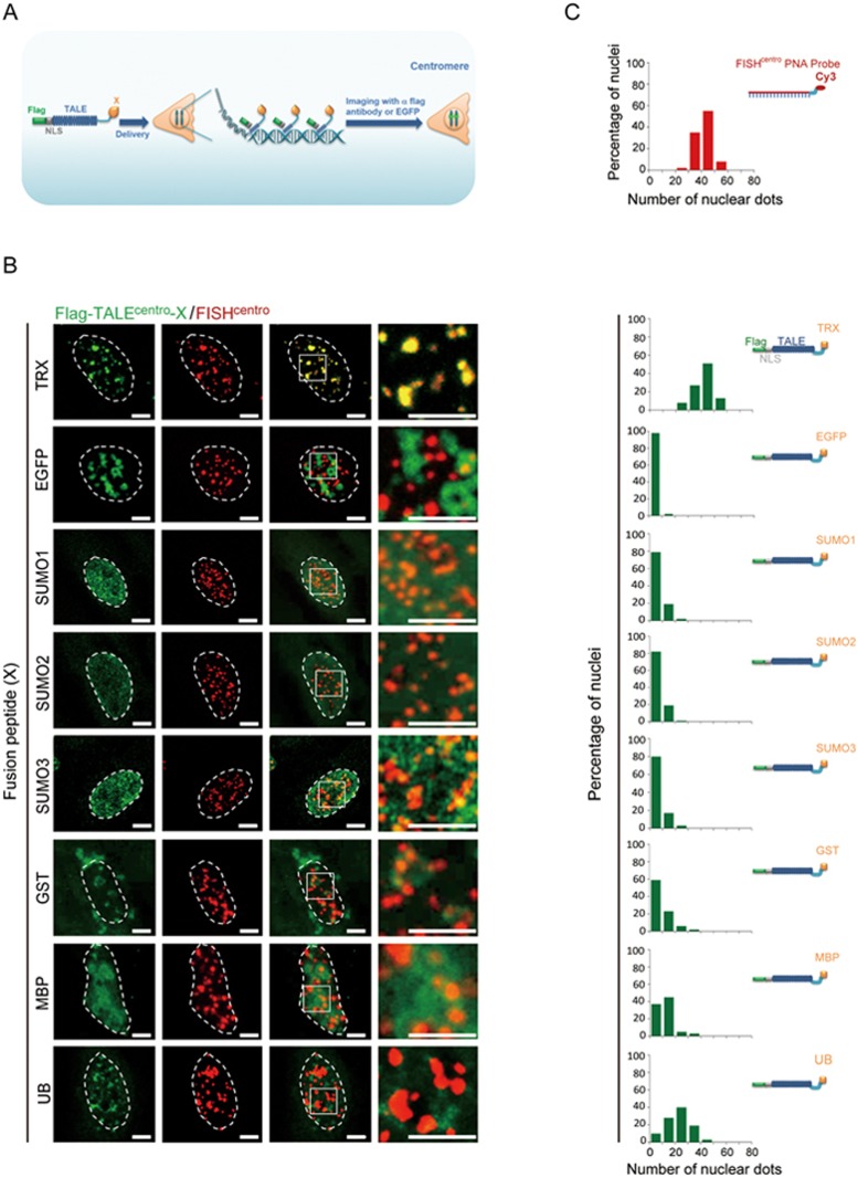
Figure 3.
Precise labeling of centromeres with TTALEs. (A) Schematic illustration of TALEs fused with various solubility-enhancing peptides (X) to label centromeres. (B) Co-localization analysis of centromeric FISH (red) and Flag-TALEcentro (green) fused with the indicated peptides in HeLa cells. Engineered TALEcentro was visualized by immunostaining with anti-Flag antibody. Representative images show precise co-localization of centromeric FISH (red) and TRX-fused TALEcentro (TTALEcentro) signals. Dashed lines indicate the nuclear boundary. Scale bars, 5 μm. (C) Histograms showing numbers of centromeric FISH- and peptide-fused TALEcentro(19 bp)-positive dots in nuclei of HeLa cells. n = 50 nuclei per group.

