FIG. 6.
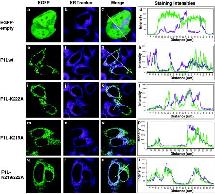
The loss of basic amino acids in the C-terminal tail of F1L results in ER localization. HeLa cells were transfected with either pEGFP-empty vector (a), pEGFP-F1Lwt (e), pEGFP-F1L-K222A (i), pEGFP-F1L-K219A (m), or pEGFP-F1L-K219/222A (q) and stained with ER-Tracker (b, f, j, n, and r). Merged images and intensity plots (c, d, g, h, k, and l) indicate that EGFP-F1Lwt and EGFP-F1L-K222A do not localize to the ER. The merge image of EGPF-F1L-K219A and ER Tracker (o) indicates partial ER localization, which is further supported by the merge intensity plot (p). The merge image of EGFP-F1L-K219/222A and ER-Tracker (s) indicates EGFP-F1L-K219/222A localizes to the ER, which is further supported by the merge intensity plot (t).

