
Figure 5.
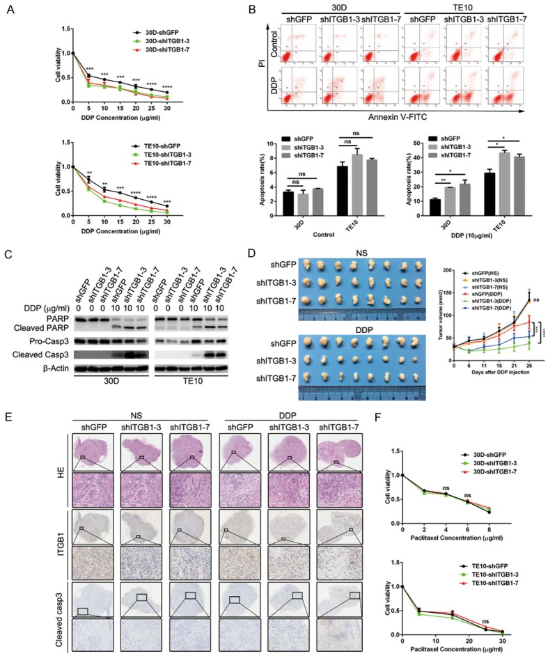
Reduced integrin β1 expression sensitizes ESCC cells to DDP administration. A: Integrin β1 deletion in 30D or TE10 cells impaired cellular viability when exposed to the indicated concentrations of DDP. B: The apoptosis rates were analyzed using flow cytometry, which showed that integrin β1 deletion promoted DDP-mediated apoptosis (10 μg/ml) in 30D or TE10 cells. C: Immunoblotting assays confirmed that DDP (10 μg/ml) treatment promoted apoptosis in integrin β1-null 30D or TE10 cells, as demonstrated by the dramatic increases in PARP and caspase-3. D: In vivo, the growth of xenografts formed by subcutaneously injected 30D cells was inhibited by 26 days of DDP treatment. Xenografts in the shITGB1-3/7 group were smaller than those in the shGFP group. E: Immunohistochemistry staining showed decreases in integrin β1 expression and increases in cleaved caspase-3 expression in the harvested xenografts. The scale bars indicate 1 mm or 2 mm in the original images and 100 μm or 200 μm in the magnified images. F: The deletion of integrin β1 did not affect the viability of 30D or TE10 cells when they were exposed to the indicated concentrations of paclitaxel.