
Figure 2.
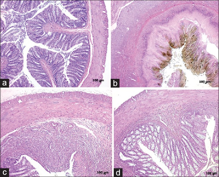
Histopathologic appearances of colon specimens: (a) Group I (control); normal colonic tissue (grade 0); (b) Group II (TNBS colitis); extensive ulceration and transmural inflammation involving the serosa (grade 5); (c) Group III (TNBS + 10 μM HNG); focal ulceration and inflammation involving the muscularis propria (grade 3); (d) Group IV (TNBS + 20 μM HNG); focal ulceration with inflammation limited to the submucosa (grade 2) (H and E; a, ×50; b, ×25; c, d, ×50)