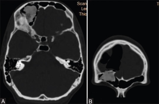
Figure 3 (A and B).

Non-enhanced computed tomography of the head (ICT 256 SLICE PHILIPS BRILLIANCE) (Bone window) axial (A) and coronal (B) images revealed homogeneous bony mass within right frontal sinus with intracranial extradural extension and associated pneumocephalus
