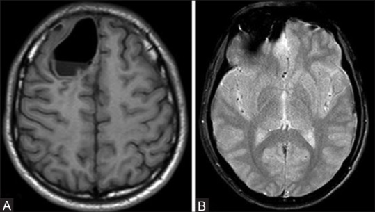
Figure 4 (A and B).

Magnetic resonance imaging of the brain (PHILIPS 1.5T) axial images reveals extra axial pneumocephalus with air fluid level appears hypointense on T1 weighted images (A), and osseous mass showing blooming on fast field echo (B)

Magnetic resonance imaging of the brain (PHILIPS 1.5T) axial images reveals extra axial pneumocephalus with air fluid level appears hypointense on T1 weighted images (A), and osseous mass showing blooming on fast field echo (B)