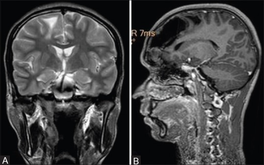
Figure 5 (A and B).

Magnetic resonance imaging of the brain T2 weighted coronal (A) demonstrating subcortical white matter edema in the right frontal lobe and sagittal T1 weighted post-contrast image (B) reveals isointense osseous lesion in right frontal sinus with associated pneumocephalus with air fluid level without any peripheral enhancement and mass effect on right frontal and high parietal lobe with surrounding edema
