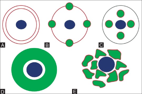
Figure 1 (A-E).

Schematic line diagram showing development of biliary tract. (A) During 8th gestational week, ductal plate (brown), becomes apparent in mesenchyme surrounding portal vein radicle (blue). (B) By 12th gestational week, remodelling starts and parts of ductal plate fuse and are reabsorbed. (C) Unfused portions constitute definitive bile ducts (green). (D) Ductal plate malformation in which continuous dilated duct encircles the portal vein radicle; and (E) interrupted circle of ectatic bile ducts
