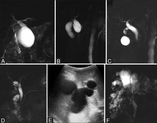
Figure 11 (A-F).

Choledochal cyst. (A) Type IA – marked cystic dilatation of entire extrahepatic bile duct (B) Type IB – focal saccular dilatation (C) Type IC –smooth fusiform dilatation of entire extrahepatic bile duct (D) Type II –discrete diverticulum arising from lateral wall of common hepatic duct (E) Type IVA –multiple sites of dilatation of both extrahepatic and intrahepatic biliary tree (F) Type V –multiple sites of saccular or cystic dilatation of only intrahepatic biliary tree (Caroli disease)
