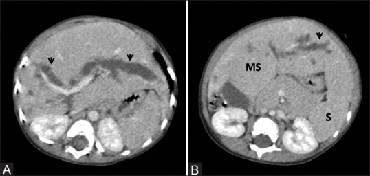
Figure 6 (A and B).

Congenital hepatic fibrosis with Caroli's disease. (A, B) Contrast-enhanced portal venous phase images depicting marked dilatation of intrahepatic biliary radilces (arrowheads) with associated findings of congenital hepatic fibrosis – atrophy of right lobe and hypertrophy of medial segment (MS) of left lobe with signs of portal hypertension, namely, splenomegaly (s)
