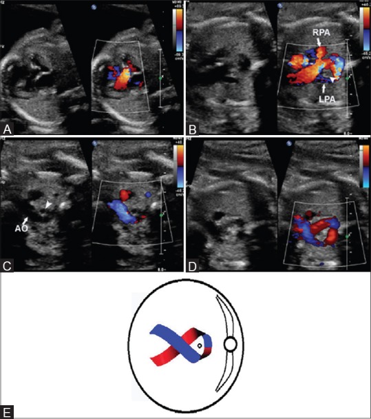
Figure 1 (A-E).

(A) Left ventricular outflow tract view showing the normal origin of aorta from the left ventricle and directed towards the right shoulder. (B) Section just cephalic to A showing the origin of the main pulmonary artery from right ventricle and extending to the right and giving off the branches of right (RPA) and left (LPA) pulmonary arteries and continues to the right of the trachea (arrow head) as ductus arteriosus. (C) Section just cephalic to B shows the aortic arch (AO) extending to the left of the trachea (arrow head). (D) Slightly oblique scan showing the aortic arch to the right and the ductus arteriosus to the left of the trachea and joining together behind the trachea giving a cross-ribbon appearance (E) Line diagram showing the cross ribbon sign
