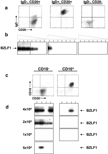
FIG. 4.
EBV replicates in the IgD− CD10− CD20+ subset of tonsil B cells. (a) T-cell-depleted tonsil lymphocytes (CD3−) were fractionated by MACS into IgD+ and IgD− fractions. The IgD− subset was further fractionated by MACS into CD20+ and CD20− fractions. The purified cells were analyzed by flow cytometry for purity (top panels). (The T-cell depletion was less efficient for this experiment than for the one shown in Fig. 2.) The CD20− fraction probably consists of contaminating T cells and macrophages but could also include plasma cells that had lost their lineage markers. (b) Cells (106) of each fraction were subjected to serial twofold dilutions, and the dilutions were tested by RT-PCR for expression of the immediate-early gene BZLF1. PCR products were identified by Southern blotting with a gene-specific probe. Tonsil 1 from Table 1 was used in this experiment. (c) CD10+ CD20+ and CD10− CD20+ tonsil lymphocytes were separated by FACS sorting. (d) The samples were serially diluted, and then multiple replicates were made for each dilution. Each sample was analyzed by RT-PCR (upper panels) for expression of the immediate-early BZLF1 gene. PCR products were identified by Southern blotting with a gene-specific probe. Tonsil 2 from Table 1 was used in this experiment.

