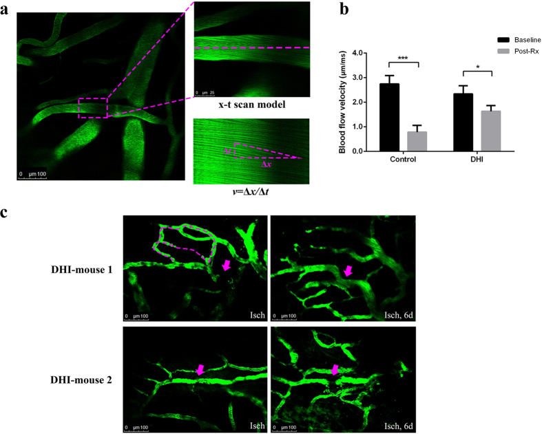
Figure 2. Effects of DHI on cerebrovascular thrombosis in mice.
A cerebral microarteriole thrombosis mouse model was established by using the two photon laser scanning microscopy. (a) Measurement of arteriole blood flow-velocity by line-scans along the longitudinal axis of target arterioles. The slopes (△x/△t) of the measurement-angles are proportional to flow velocity. (b) Arteriole with diameter of 18 ± 3 μm and 3 ± 1 μm/ms in blood velocity was selected as the target vessel for occlusion. DHI was injected after the thrombus formation. After 30 min, the blood flow velocity was calculated to evaluate the effect of DHI. The graph depicts the mean ± standard deviation (n = 5). * P < 0.005 and *** P < 0.0001 as compared with the baseline in each group. (c) New branches of arterioles appeared six days after the proximal vessels occluded in two mice of DHI group. Isch, after thrombus induction, before DHI treatment; Post Rx, after administration of DHI. Magenta dashed: location of disappeared vessels. Magenta arrows: occlusion site through laser irradiation.

