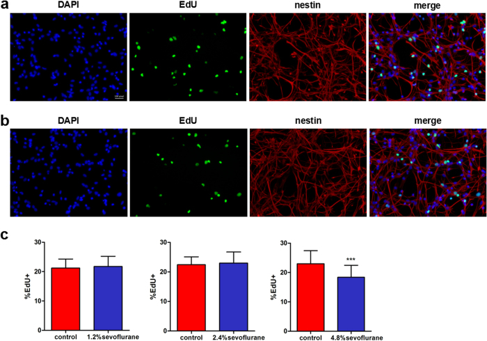
Figure 4. Cell proliferation was assessed immunocytochemically using the EdU incorporation assay immediately after anesthesia.
(a) Immunofluorescence images were obtained by the EdU incorporation (green, Alexa Fluor 488) assay, DAPI (blue) nuclear staining for EdU-positive cells, and Alexa Fluor 555-stained (pink, Alexa Fluor 555) for nestin-marked cells and then the images were merged. (b) Immunofluorescence images from FNSCs exposed to 4.8% sevoflurane for 6 h. (c) Statistical results of EdU-positive cell ratio in FNSCs exposed to different concentrations of sevoflurane (1.2%, 2.4%, and 4.8%) for 6 h. Data were obtained from at least three separate cultures, ***P < 0.001 versus control group.

