Figure 1.
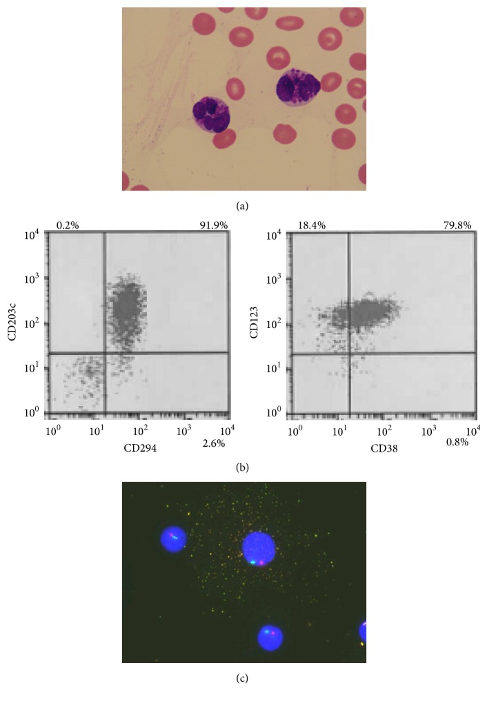
The characterization of basophils in peripheral blood. (a) Peripheral blood smear showed some medium- to large-sized abnormal cells with lobulated nucleus, and many basophilic granules were observed (May-Giemsa staining, original magnification, ×1000). (b) Flow cytometric analysis showed that abnormal cells were positive for CD38, CD123, CD203c, and CD294, which was consistent with basophils. (c) Dual-color FISH analysis using chromosome 7 probes showed that abnormal cell with many granules contained one red signal and one green signal, which indicated monosomy 7 (D7S486 probe, red signal; D7Z1 probe, green signal). Other cells without any granules also showed monosomy 7.

