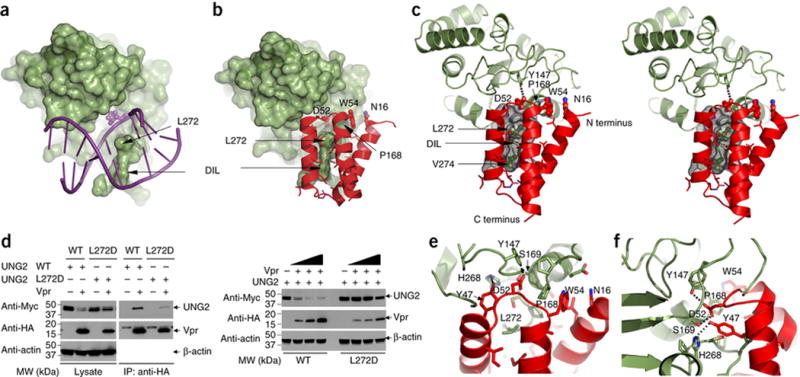
Figure 2. Vpr employs structural mimicry and engages UNG2 residues that are also used in DNA binding.

(A) Structure of the UNG2 (green) - DNA (purple) complex (PDB:id 2OYT,)42). UNG2 is depicted in surface representation and the double stranded DNA in stick representation. The flipped-out abasic sugar inside UNG2’s active site pocket is shown in space filling representation. The Leu272 residue and the DNA intercalation loop (DIL) are labeled by arrows.
(B) Structure of the UNG2 (green) - Vpr (red) portion of the DDB1-DCAF1-Vpr-UNG2 complex. UNG2 is depicted in surface representation and Vpr in ribbon representation. Several side chains of Vpr that are interacting with UNG2 are shown in ball and stick representation and labeled with residue name and number.
(C) Stereo-view of the contact region between UNG2 (green) and Vpr (red), illustrating details of the interaction. Leu272 and Val274 of UNG2 are buried inside Vpr’s hydrophobic cleft (rendered in gray surface representation). Asp52 of Vpr (ball and stick representation) is located inside UNG2’s active site pocket, forming a hydrogen bond with Tyr147 of UNG2. Trp54 of Vpr (ball and stick representation) is in van der Waals contact with Pro168 of UNG2 and Asn16 of Vpr is stacked onto the Trp54 side chain.
(D) Co-immunoprecipitation experiments to assess the interaction between UNG2 and Vpr (left panels). UNG2 WT or Leu272Asp was transfected into HEK293 cells with increasing amounts of Vpr (right panels).
(E–F) Two expanded views of Vpr’s insert loop contacts with the active site pocket of UNG2.
