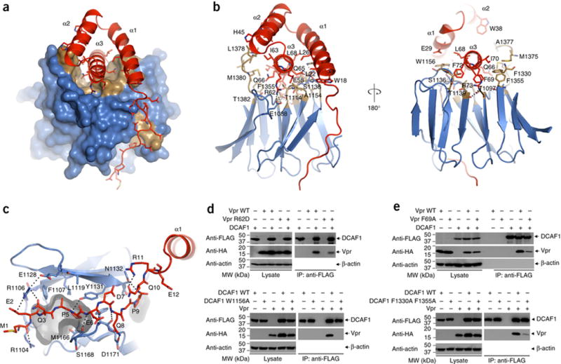
Figure 3. Interface between DCAF1 (blue and gold) - Vpr (red) in the DDB1-DCAF1-Vpr-UNG2 complex.

(A) Surface (DCAF1) and ribbon (Vpr) representations of the DCAF1-Vpr portion of the DDB1-DCAF1-Vpr-UNG2 complex structure. The hydrophobic cleft in the rim of the DCAF1 WD40 propeller, which accommodates the a3 helix of Vpr is shown in gold.
(B) Two views of DCAF1-Vpr interactions, illustrating details of contact residues. Pertinent side chains are shown in stick representation. Vpr is shown in red, and DCAF1 is in gold (hydrophobic) and blue (polar).
(C) Detailed view of the interaction between the N-terminal tail of Vpr and residues of DCAF1 at the edges of two propeller blades. A small hydrophobic pocket, accommodating Pro5 of Vpr is rendered in gray.
(D) – (E) Co-immunoprecipitation experiments to examine the effect of selected mutants on Vpr-DCAF1 binding.
