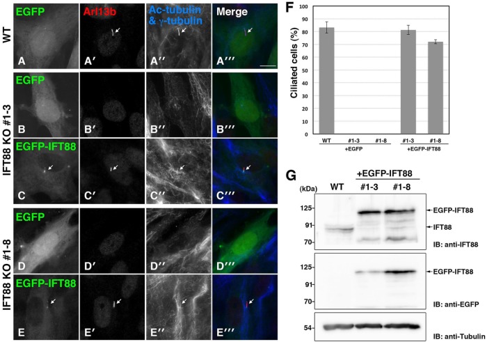
FIGURE 3:
Rescue experiments of IFT88-KO cell lines. WT RPE1 cells (A) and IFT88-KO cell lines #1-3 (B, C) and #1-8 (D, E) were transfected with pEBMulti-Ble-EGFP (A, B, D) or pEBMulti-Ble-EGFP-IFT88 (C, E). The transfected cells were cultured under serum-starved conditions and immunostained for Arl13b (A’–E’), Ac-tubulin, and γ-tubulin (A”–E”). Merged images are shown in A”’–E”’. Arrows indicate primary cilia. Scale bar, 10 μm. WT cells and IFT88-KO cells exogenously expressing EGFP-IFT88 were ciliated, whereas IFT88-KO cells expressing EGFP were not. (F) Ciliated cells in the experiments shown in A–E were counted, and percentages of ciliated cells are represented as bar graphs. Values are means ± SE (error bars) of three independent experiments. In each experiment, 40–80 cells with EGFP signals were examined. (G) Confirmation of exogenously expressed EGFP-IFT88 in IFT88-KO cell lines by immunoblotting. Lysates from WT RPE1 cells and from IFT88-KO clones #1-3 and 1-8 expressing EGFP-IFT88 were immunoblotted with antibodies against IFT88 and GFP. Tubulin was analyzed as a loading control.

